
Diagnostic imaging

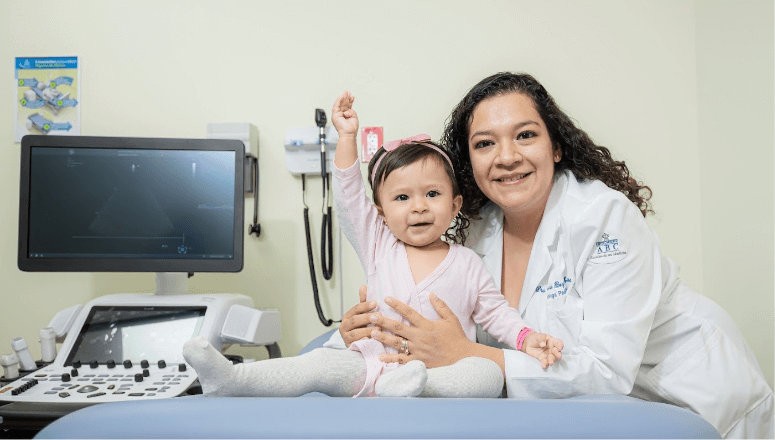
Your child's heart in the best hands
ABC Medical Center and Kardias have jointly developed a highly specialized program for the care of congenital cardiovascular diseases in pediatric patients.
Diagnostic imaging
Congenital heart diseases comprise a wide spectrum of heart malformations, which can be very simple or complex defects. The primary imaging tools are chest x-ray and cardiac ultrasound. When it is necessary to better define the morphology and function of the heart disease, pediatric cardiologists use other imaging methods with higher resolution, which are classified as invasive methods (cardiac catheterization) and non-invasive methods (MRI and CT Scan).
How to detect a congenital heart disease?
Congenital heart disease can be detected in the womb or when the baby is born. In this video you will find the three key moments in which it can be detected and what you can do at each stage or moment.
Some warning signs are chest pain, strong palpitations, fatigue, excessive sweating, and growth problems. See your doctor if you detect any warning signs. From the uterus to childhood the specialists at the ABC–Kardias Pediatric Heart Center can help you make the best decision for your child’s well-being.
Computed tomography
It is a technique that uses a computer to create cross-sectional images of the heart. The images are taken in the imager, which is a large tube-shaped X-ray machine where the child is introduced and radiographic images are obtained as if they were slides or slices, a contrast medium can be administered to see the vessels and heart chambers (CT angiography). From these images, the computer can reconstruct a detailed representation of the heart, even in 3D.
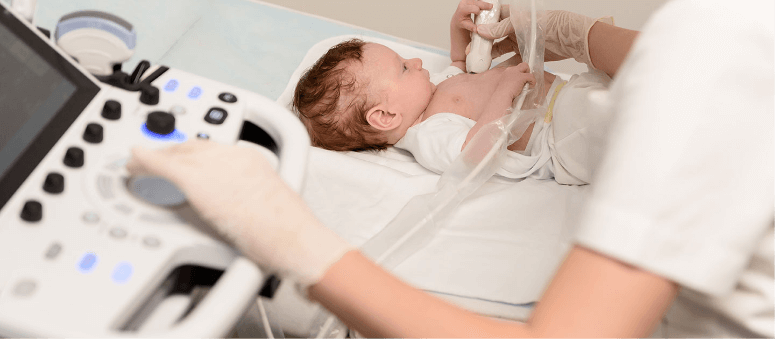
Pediatric echocardiography
We have everything necessary to perform pediatric echocardiography using sound waves to create images of the heart. Through these images, the specialist identifies and diagnoses the disease that the patient is suffering from, and proposes the treatment to follow. The echocardiographer cardiologist works hand in hand with the cardiovascular surgeon before and during the procedure and will monitor your child while they remain in the PCICU.

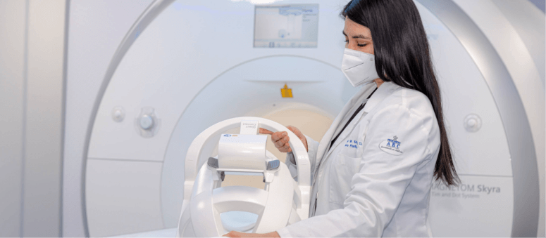
MRI
It is an imaging test that allows clear images of the heart to be obtained without the need for a catheterization procedure and without using radiation. This machine is very similar to the imager, it has the shape of a long, narrow tube, inside which the child is placed and is surrounded by a magnetic field that interacts with the elements of their body, most of which has a magnetic charge. Using a computer, the magnetic signals it produces are read and converted into an image that can be seen on a screen. A contrast medium can also be administered to enhance the vessels (MR angiography) and better define the heart structures. It is a useful test to evaluate cardiac function and define deep structures.
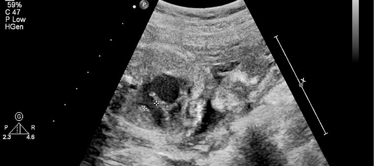
Fetal cardiac ultrasound
Most congenital heart malformations can be detected in the fetal stage. The fetal cardiac ultrasound provides an accurate diagnosis for the appropriate treatment for each patient at the time of birth or during the first months of life, if necessary.
Where to Find Us

Campus Observatorio
Sur 136 No. 116, Col. Las Américas, Álvaro Obregón, 01120, Cd. de México.

Campus Santa Fe
Av. Carlos Graef Fernández 154, Col. Santa Fe, Cuajimalpa, 05300, Cd. de México.
