Contact us
Precision Medicine
-
Healthcare Professionals
Healthcare Professionals
Credentialing Requirements
- About ABC
-
Inclusive Health
- ABC Talent
- ABC Foundation I.A.P
- Contact
- ES
What is three-dimensional echocardiogram?
10 November 2025
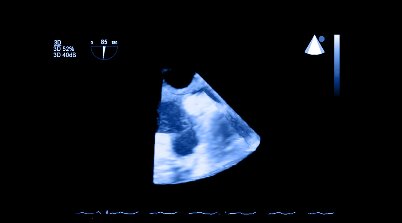
A three-dimensional echocardiogram, or 3D echocardiogram, is a test that uses ultrasound waves to obtain three-dimensional images. This allows for a clearer and more precise visualization of different characteristics of the heart’s anatomy, including its shape, strength, size, and movement.
What Is a Three-Dimensional Echocardiogram?
A three-dimensional echocardiogram is an advanced cardiac imaging technique that allows for the heart to be visualized in three dimensions and in real-time. Unlike a traditional two-dimensional echocardiogram, this technique offers a volumetric representation of the heart, which facilitates a more precise evaluation of its anatomy and function.
Although both this study and the conventional echo use ultrasound, in the three-dimensional one, with the support of special software and transducers, the images are reconstructed in 3D. This is especially useful for studying the valves, chambers, and internal structures in greater detail.
A three-dimensional echocardiogram has become a fundamental tool in cardiology, especially for the planning of heart surgeries, the monitoring of valve diseases, and for the evaluation of implantable devices like pacemakers or prostheses.
Thanks to its ability to show the heart from different angles and in motion, clinical decisions can be made, reducing the need for invasive studies.
How Is a Three-Dimensional Echocardiogram Performed?
Performing a three-dimensional echocardiogram begins in a similar way to a conventional echocardiogram. The patient lies down on a stretcher, and a conductive gel is applied to their chest to facilitate the transmission of ultrasound waves.
Subsequently, the specialist places a transducer on the chest, which emits and receives the necessary signals to form images of the heart. In the case of a 3D echocardiogram, more advanced transducers are used, as well as specialized software to capture multiple planes simultaneously. This is what allows for the reconstruction of a three-dimensional image in real-time.
The entire procedure usually lasts from 30 to 60 minutes and generally does not require any special preparation, except in the case of the transesophageal study.
It is a safe, well-tolerated, and very valuable study for obtaining highly accurate anatomical and functional information.

After the Three-Dimensional Echocardiogram Study
Depending on each case, the analysis can be done in real-time during the study or afterward to review the images from different angles or to make specific measurements.
The result of the study is delivered shortly, accompanied by a detailed report that the treating physician will use to confirm a diagnosis or adjust the treatment plan.
With this information, various steps can be taken. If the study shows that the heart is functioning normally, no additional treatment may be necessary.
But in case of detecting abnormalities such as a damaged valve, a structural defect, or heart failure, the doctor may order more studies, start a medical treatment, or evaluate a surgical intervention.
This type of study is usually requested in situations where a detailed visualization of the heart is required, such as in the evaluation of valve diseases, monitoring of congenital heart diseases, planning of heart surgery, or placement of intracardiac devices.
It is also very useful for assessing the function of the left ventricle after a heart attack or in patients with heart failure.
In general, this study is reserved for cases where the two-dimensional echocardiogram does not offer enough information or when a more complete image is needed for a medical decision to be made.
At the Cardiovascular Diagnostics area at ABC Medical Center, we can provide you with specialized care. Contact us!
Fuentes:
How can we help you?

Ricardo Ostos
Content CreatorRicardo can convey complex medical information in an accessible and friendly way so that all of our patients can understand and benefit from it. In addition, he has an empathetic approach, offering information and practical advice that really makes a difference in people's lives. #lifebringsustogether.
Learn more about Ricardo on LinkedIn
Pay in interest-free monthly installments in Specialty Centers, Check Ups, Diagnostic Tests, and Hospitalization
Get from 3 to 9 interest-free installments* with American Express or 6 installments* when paying with Banamex, BBVA, HSBC, Santander or 12 installments*
when paying with Banamex.
Privacy Overview
Error: Contact form not found.
We help you
Send us your request and we will forward it to our specialists. We will get in touch with you very soon.
If you have preferred times to receive our call, please indicate them in your message.
Thank you for contacting us!
Interest-free
months in:
Interest-free
months in:
Specialty Centers
Diagnostic Studies
Check-ups
Hospitalization1
Choose from3 to 9 months when paying with American Express cards 2. Or
6 months when paying with your credit card3 Banamex, BBVA Bancomer, HSBC, Santander.
Or 12 months exclusively when paying with Banamex3
Valid until December 31, 2025. Promotions not cumulative. Subject to restrictions 1. In hospitalization, medical fees are not included. 2. Minimum amount: $1,500 for 3 to 6 months and $3,000 for 7 to 9 months 3. Minimum amount $1,500. (Cards issued abroad are not eligible).
Comparison of COVID-19 vaccines
Pfizer-
BioNTech
Pfizer-BioNTech
What is its effectiveness and what does it refer to?
Vaccine type: mRNA
Effectiveness: 95% after the second dose in the prevention of symptomatic COVID-19.
No Does not contain egg, latex, or preservatives.
How many doses are needed?
Two doses are needed, at least 21 days apart (or up to six weeks apart, if necessary).
Who should or shouldn’t get the vaccine?
People who should receive the vaccine are those over 16 years old.
People who should not receive the vaccine are those who have a history of anaphylactic shock (severe allergy) or who are allergic to any component of this vaccine such as polyethylene glycol (PEG) or polysorbate.
What are the possible side effects of the vaccine?
Pain where the injection was given, fatigue, headache, muscle pain, chills, joint pain, fever, nausea, malaise, and swollen lymph nodes.
How long will it take for me to be protected and what does it protect me from?
After 14 days of having the complete scheme (after the administration of the 2nd dose), the protection period is still under study. It protects us from serious COVID-19 or requiring hospitalization.
Moderna
What is its effectiveness and what does it refer to?
Vaccine type: mRNA
Effectiveness: 94.5% after the second dose in the prevention of symptomatic COVID-19.
Does not contain egg, latex, or preservatives.
How many doses are needed?
Two doses are needed, at least 28 days apart (or up to six weeks apart, if necessary).
Who should or shouldn’t get the vaccine?
People who should receive the vaccine are those over 18 years old.
People who should not receive the vaccine are those who have a history of anaphylactic shock (severe allergy) or who are allergic to any component of this vaccine.
What are the possible side effects of the vaccine?
Pain where the injection was given, fatigue, headache, muscle pain, chills, joint pain, fever, nausea, and swollen lymph nodes in the arm in which you received the injection.
How long will it take for me to be protected and what does it protect me from?
After 14 days of having the complete scheme (after the administration of the 2nd dose), the protection period is still under study. It protects us from serious COVID-19 or requiring hospitalization.
Janssen/
Johnson
& Johnson
Janssen/ Johnson & Johnson
What is its effectiveness and what does it refer to?
Vector-based vaccine.
Effectiveness: 72.0% in the prevention of symptomatic COVID-19.
85% in the prevention of severe COVID-19.
Does not contain egg, latex, or preservatives./strong>
How many doses are needed?
Only one dose in needed.
Who should or shouldn’t get the vaccine?
People who should receive the vaccine are those over 18 years old.
People who should not receive the vaccine are those who have a history of anaphylactic shock (severe allergy) or who are allergic to any component of this vaccine.
What are the possible side effects of the vaccine?
Pain where the injection was given, headache, fatigue, muscle pain, chills, fever, and nausea.
How long will it take for me to be protected and what does it protect me from?
After 28 days of having the complete scheme (the last dose applied), the protection period is still under study. It protects us from 85% serious COVID-19 or requiring hospitalization.
AstraZeneca
and
Oxford
University
AstraZeneca and Oxford University
What is its effectiveness and what does it refer to?
Adenovirus vector-based vaccine.
Effectiveness: 82% after the second dose in the prevention of symptomatic COVID-19.
How many doses are needed?
Two doses are needed, at least 56 days apart (or up to 84 days apart, if necessary).
Who should or shouldn’t get the vaccine?
People who should receive the vaccine are those over 18 years old.
People who should not receive the vaccine are those who have a history of anaphylactic shock (severe allergy) or who are allergic to any component of this vaccine.
What are the possible side effects of the vaccine?
Pain where the injection was given, fatigue, headache, myalgia, arthralgia, and fever, which were mild to moderate in intensity and disappeared within 48 hours of vaccination.
How long will it take for me to be protected and what does it protect me from?
After 14 days of having the complete scheme (after the administration of the 2nd dose), the protection period is still under study. It protects us from serious COVID-19 or requiring hospitalization.
Sputnik V
What is its effectiveness and what does it refer to?
Adenovirus vector-based vaccine.
Effectiveness: 92% after the second dose in the prevention of symptomatic COVID-19.
How many doses are needed?
Two doses are needed, at least 21 days apart (or up to six weeks apart, if necessary).
Who should or shouldn’t get the vaccine?
People who should receive the vaccine are those over 18 years old.
People who should not receive the vaccine are those who have a history of anaphylactic shock (severe allergy) or who are allergic to any component of this vaccine.
What are the possible side effects of the vaccine?
Pain where the injection was given, fatigue, headache, myalgia, arthralgia, and fever, which were mild to moderate in intensity and disappeared within 48 hours of vaccination.
How long will it take for me to be protected and what does it protect me from?
After 14 days of having the complete scheme (after the administration of the 2nd dose), the protection period is still under study. It protects us from serious COVID-19 or requiring hospitalization.
Anti-Herpes Zoster
Herpes zoster is a painful, burning rash. It usually appears on one part of the body and can last for several weeks. It can cause long-lasting severe pain and scarring. Bacterial skin infections, weakness, muscle paralysis, hearing or vision loss may occur less frequently. Herpes zoster is caused by the same virus that causes chickenpox. After you have had chickenpox, the virus that caused it remains in the body of nerve cells. Sometimes after many years, the virus becomes active again and causes herpes zoster.
Vaccination is indicated in the following cases:
- Immunization of patients from 50 years of age for the prevention of herpes zoster and post-herpetic neuralgia (PHN), reduce pain associated with acute or chronic herpes zoster.
Scheme type:
- Single dose.
Rabies
Human rabies is a viral disease transmitted by the bite of an infected animal. It is characterized by acute encephalomyelitis (an aggressive response of the immune system that destroys the myelin layer of the nerves and alters its function at the level of the brain or spinal cord).
Vaccination is indicated in the following cases:
- Prevention of rabies in subjects exposed to risk of contamination. It is recommended for certain international travelers, based on the occurrence of animal rabies in the destination country.
Scheme type:
There are two types.
1. Pre-exposure scheme, consists of three doses of rabies vaccine:
- First dose, on the chosen date.
- Second dose 7 days after the first dose.
- Third dose 28 days after the first dose.
2. Post-exposure scheme, people not vaccinated against rabies, consists of five doses of rabies vaccine.
- First dose, on the date indicated.
- Second dose 3 days after the first dose.
- Third dose 7 days after the first dose.
- Fourth dose 14 days after the first dose.
- Fifth dose 28 days after the first dose.
* If the individual continues to be at risk of exposure to the disease, revaccination should be considered.
Pneumococcal vaccines
Pneumococcal disease can cause serious infections in the lungs (pneumonia), the bloodstream (bacteremia), and the lining of the brain and spinal cord (meningitis).
Two vaccines help prevent pneumococcal disease:
- Pneumoconjugate 13 (pneumococcal conjugate vaccine)
- Pneumococcal 23 (pneumococcal polysaccharide vaccine)
Vaccination is indicated in the following cases:
- Active immunization for the prevention of invasive disease caused by Streptococcus pneumoniae in adults 65 years of age and older.
Scheme type:
- *Two pneumococcal vaccines are recommended for all adults 65 years of age or older.
*One dose of Pneumococcal 13 vaccine should be given first, followed by one dose of Pneumococcal 23 vaccine, depending on your age and health.
