
Contact us
Precision Medicine
What is cardiac mri?
10 November 2025
A cardiac MRI is a study that allows for the acquisition of high-resolution images of different areas of the heart to precisely evaluate its function.
What Is a Magnetic Resonance Imaging?
Magnetic resonance imaging works by using magnetic fields and the emission of radio waves, which allows for the acquisition of detailed images without being invasive to the patient or using radiation.
A cardiac MRI is commonly used to assess the size, shape, and function of the heart and to detect different conditions such as coronary artery abnormalities, congenital heart problems, or conditions located in a heart muscle.
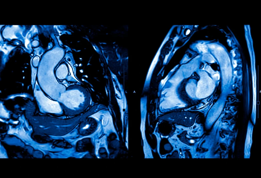
A cardiac MRI is a non-invasive imaging study for which the patient lies on a stretcher and enters a closed tunnel of the equipment. Electrodes are placed on the chest to synchronize the images with the heart rhythm, which is known as electrocardiographic gating. In some cases, an intravenous contrast medium may be administered.
The procedure usually lasts between 30 and 60 minutes, depending on the type of study and the protocol used.
Cardiac MRI Protocol
The cardiac MRI protocol refers to the set of specific sequences and techniques used during the study to respond to a specific clinical suspicion.
Therefore, there is no single universal protocol, as it is adapted according to the condition to be evaluated, such as ischemic heart disease, cardiomyopathies, or congenital heart diseases, to name a few examples.
Almost all studies include functional images that allow for the observation of the heart’s movement in real-time and the evaluation of ventricular contractility.
In addition to this functional analysis, the protocol usually includes tissue characterization sequences, such as T1 and T2-weighted images, parametric maps, and late gadolinium enhancement. These techniques are fundamental for detecting inflammation, edema, fibrosis, or iron overload in the myocardium.
Each protocol is designed to obtain the maximum diagnostic information with the least possible discomfort for the patient.
Interpreting a Cardiac MRI
The interpretation of a cardiac MRI requires a detailed analysis by a medical specialist in cardiovascular imaging.
It begins with the evaluation of the moving images, where the systolic and diastolic function of the ventricles is analyzed, and volumes, ejection fraction, myocardial mass, and abnormalities in wall movement are calculated.
Subsequently, the tissue characterization sequences are examined to detect signs of edema, fibrosis, fatty infiltration, iron overload, or necrosis.
These findings are interpreted in the clinical context of each patient, as many imaging patterns are characteristic of specific conditions.
At the Cardiovascular Diagnostics area at ABC Medical Center, we can provide you with specialized care. Contact us!
Fuentes:
How can we help you?

Ricardo Ostos
Content CreatorLearn more about Ricardo on LinkedIn
The dissemination of the content of this material is for informational purposes only and does not replace, under any circumstance or condition, a consultation with a specialist doctor, for which the ABC Medical Center is not responsible for the different use that may be given to it. If you require more information related to the subject, we suggest you contact the specialist doctor you trust directly.
