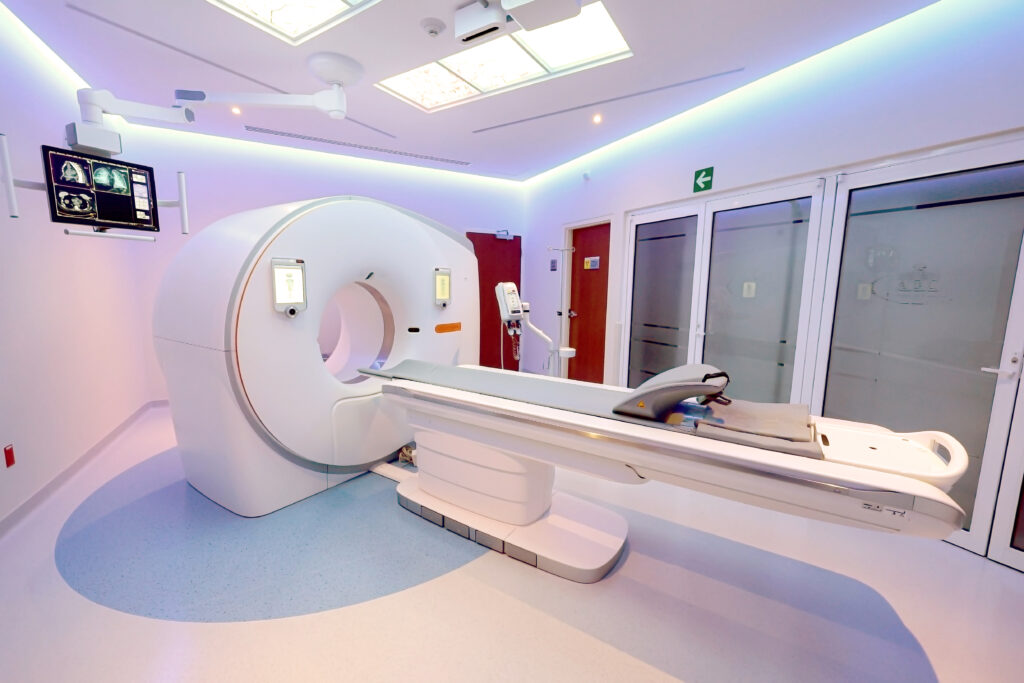
At the Department of Radiology and Molecular Imaging at Centro Médico ABC, we adopt the world’s best practices. That is why we are taking image quality to the next level with our new dual-energy CT scanners, enabling high-precision diagnoses and personalized care for every patient.
These new high-precision CT scanners allow for:
- Significant radiation dose reduction of up to 50%.
- Automatic radiation dose adjustment based on the patient’s body type and the body region being examined.
- Reduced contrast media dosage, which is particularly beneficial for patients with kidney damage, older adults, individuals with chronic conditions, metabolic disorders, or overweight—making procedures gentler on the kidneys.
- High-resolution imaging with low radiation doses.
- Morphological and functional evaluation (perfusion: blood flow to an organ), enabling tumor detection and assessment of cancer treatment response.
These new high-precision CT scanners allow for:
- Significant radiation dose reduction of up to 50%.
- Automatic radiation dose adjustment based on the patient’s body type and the body region being examined.
- Reduced contrast media dosage, which is particularly beneficial for patients with kidney damage, older adults, individuals with chronic conditions, metabolic disorders, or overweight—making procedures gentler on the kidneys.
- High-resolution imaging with low radiation doses.
- Morphological and functional evaluation (perfusion: blood flow to an organ), enabling tumor detection and assessment of cancer treatment response.
- Reduction of image artifacts caused by patient movement (especially in pediatric, emergency, intensive care, or hospitalized patients) due to rapid scan acquisition, as well as artifacts caused by metallic devices or prostheses thanks to specialized filters.
- Safer interventional studies with a fluoro-CT program that enables real-time procedure guidance.
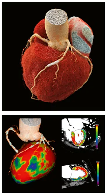
- High-definition coronary evaluation and myocardial perfusion in 10 minutes.
- Coronary anatomy evaluation in just 2 heartbeats, 1.2 seconds, and with less than 2 mSv of radiation.
- Cardiac and vascular studies with over 50% less radiation than conventional CT scanners and 40% less contrast agent, utilizing ultra-fast acquisition protocols (“Turbo Flash Scan”).
- Reduction of image artifacts caused by patient movement (especially in pediatric, emergency, intensive care, or hospitalized patients) due to rapid scan acquisition, as well as artifacts caused by metallic devices or prostheses thanks to specialized filters.
- Safer interventional studies with a fluoro-CT program that enables real-time procedure guidance.
- High-definition coronary evaluation and myocardial perfusion in 10 minutes.
- Coronary anatomy evaluation in just 2 heartbeats, 1.2 seconds, and with less than 2 mSv of radiation.
- Cardiac and vascular studies with over 50% less radiation than conventional CT scanners and 40% less contrast agent, utilizing ultra-fast acquisition protocols (“Turbo Flash Scan”).
Applications of Dual-Energy CT Scanners
Pediatrics
- 50% reduction in radiation dose.
- Faster image acquisition to prevent patient movement from altering results.
- Broader coverage area for faster study completion.
- Generation of iodine maps that indirectly reflect pulmonary blood volume.
- Organ perfusion assessment (brain, lungs, liver, kidneys).
- Cinematic rendering (the highest-definition 3D reconstruction available).
- Morphological analysis of the heart and major blood vessels in the chest and abdomen.
- Greater patient positioning accuracy for study acquisition (3D camera).
Oncology
- 50% reduction in radiation dose.
- Reduced contrast media dosage.
- Perfusion studies of tumor lesions in solid organs (lung, liver, brain, kidneys) for treatment planning and response assessment.
- Interventional procedures (biopsies) guided by real-time fluoroscopy, facilitating lesion localization and increasing biopsy precision.
- Cinematic rendering (the highest-definition 3D reconstruction available).
- Vascular evaluation.
- Greater patient positioning accuracy for study acquisition (3D camera).
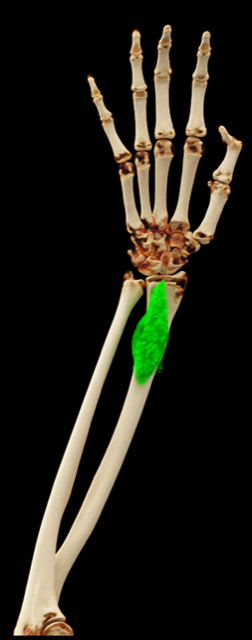
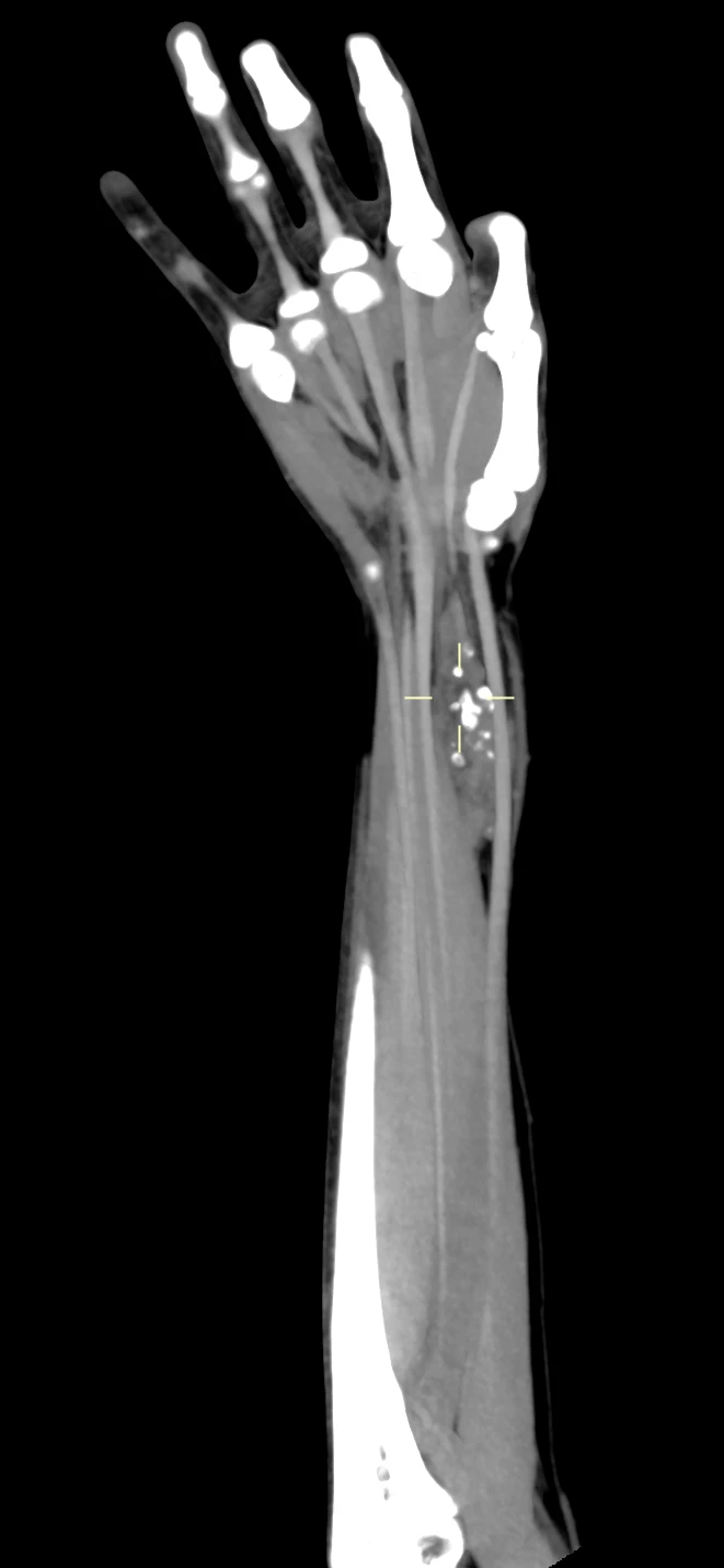
Multiplanar reconstruction of the hand and forearm showing a tumor in the distal forearm.
Orthopedics and Traumatology
- 50% reduction in radiation dose.
- Reduced contrast media dosage.
- Significant reduction of artifacts in patients with osteosynthesis hardware or metallic prostheses, allowing proper evaluation of surrounding structures.
- Detection of bone marrow edema.
- Mapping of joint inflammatory processes (e.g., rheumatoid arthritis, gout).
- Cinematic rendering (the highest-definition 3D reconstruction available).
- Interventional procedures (biopsies) guided by real-time fluoroscopy, facilitating lesion localization to increase biopsy precision.
- Greater patient positioning accuracy for study acquisition (3D camera).
- Faster acquisition speed for polytrauma patients.
- Planar rib analysis (rib unfolding/flattening to rule out fractures).
- Broader coverage area allowing for faster study acquisition.
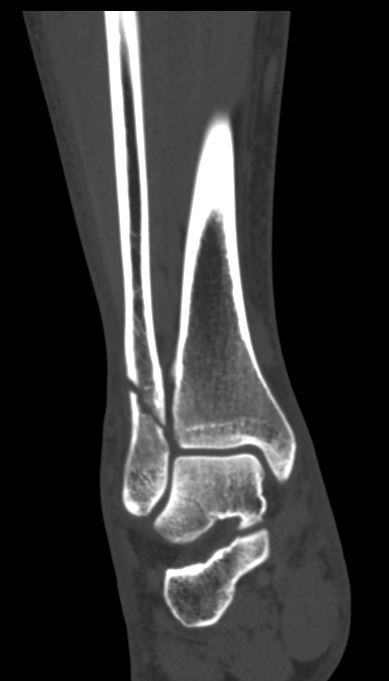
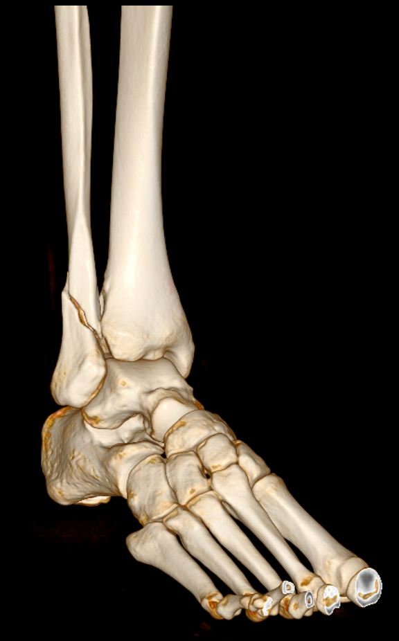
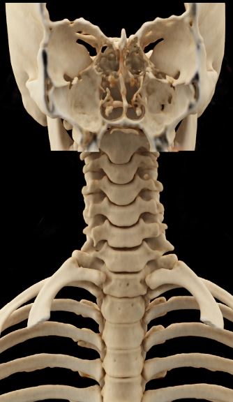
Fibula fracture in two different views and Three-dimensional image of the cervical spine (Photo 3).
Neurology
- 50% reduction in radiation dose.
- Reduced contrast media dosage.
- Brain perfusion with automated flow analysis to evaluate the ischemic penumbra in stroke patients.
- Perfusion imaging of brain tumor lesions.
- Broader coverage area allowing for faster study acquisition, particularly in cerebral vascular structures.
- Greater patient positioning accuracy for study acquisition (3D camera).
- Enhanced definition in thin slices for the ear and skull base.
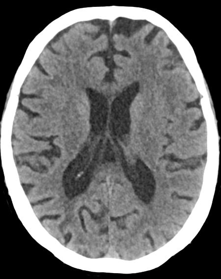
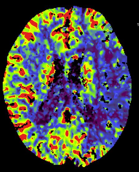
In stroke patients, applying the perfusion protocol shows the ischemic zone (Photo 2) in the left cerebral hemisphere.
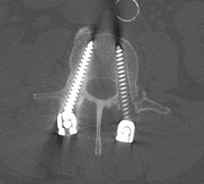
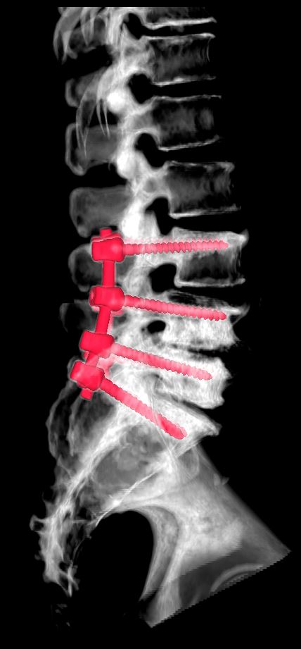
Spinal instrumentation with plates and screws.
Pulmonology
- 50% reduction in radiation dose.
- Reduced contrast media dosage.
- Pulmonary perfusion to evaluate pulmonary embolism.
- AI-driven pneumonia analysis to assess tissue involvement (volume, location, etc.).
- Broader coverage area allowing for faster study acquisition, particularly in vascular structures.
- Greater patient positioning accuracy for study acquisition (3D camera).
- Pulmonary volume assessment in patients with emphysema.
- Cinematic 3D airway reconstruction for diagnosis and treatment planning.
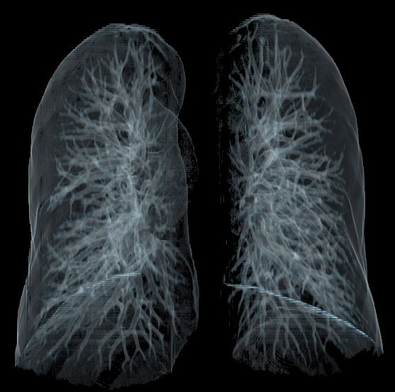
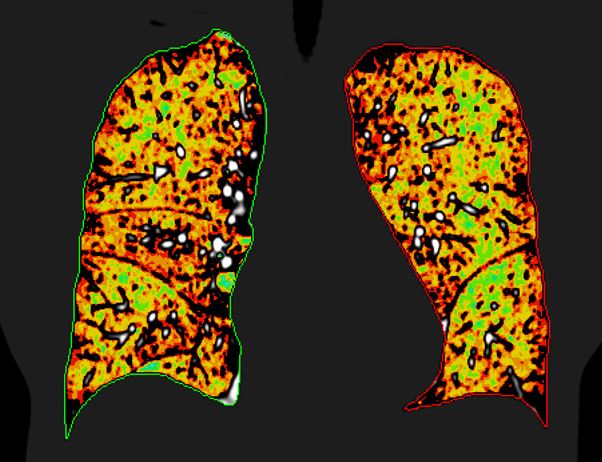
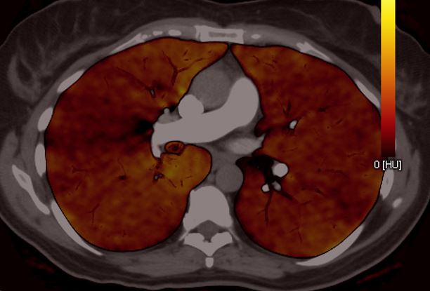
Lung images using different protocols.
Urology
- 50% reduction in radiation dose.
- Reduced contrast media dosage.
- Mineral content evaluation in stone disease (urolithiasis).
- Renal perfusion for tumor lesion evaluation.
- Broader coverage area allowing for faster study acquisition, particularly in vascular structures.
- Cinematic 3D airway reconstruction of the urinary tract for diagnosis and treatment planning.
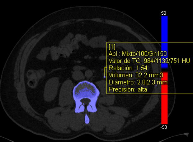
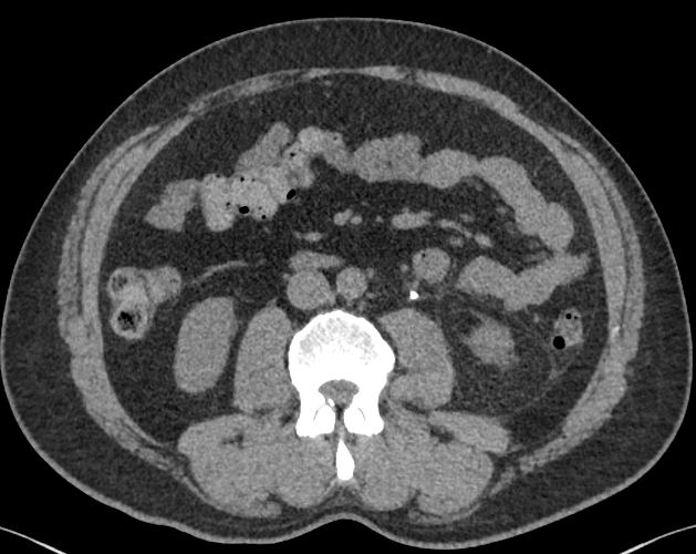
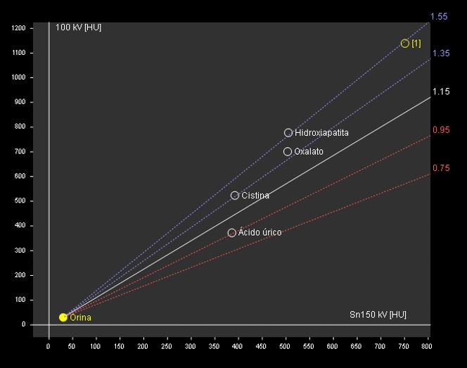
Software program that helps determine the composition of a urinary stone.
Cardiology
- 50% reduction in radiation dose.
- Reduced contrast media dosage.
- Broader coverage area allowing for faster study acquisition, particularly in vascular structures.
- CT cardiac perfusion.
- Enhanced definition of coronary arteries with reduced acquisition time, contrast dose, and radiation dose.
- Treatment planning for TAVI procedures (Transcatheter Aortic Valve Implantation).
- Cinematic 3D reconstruction of the heart and coronary arteries.
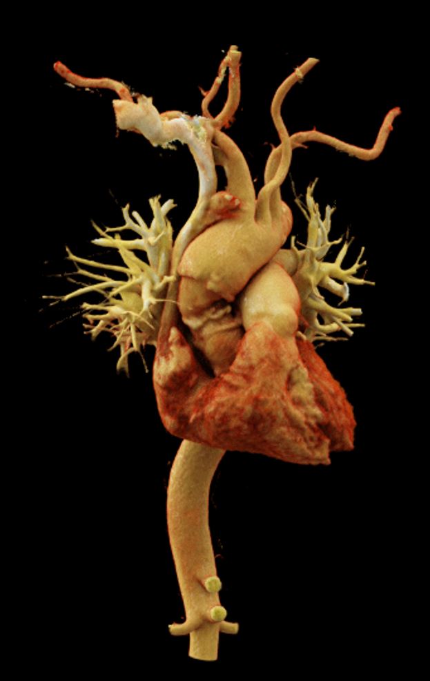
Three-dimensional image of the heart and aorta.
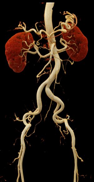
Abdominal aorta and its branches.
At the Department of Radiology and Molecular Imaging at Centro Médico ABC, we offer specialized care. Contact us today!
Fuentes
Dr. Marco Antonio Téliz Meneses – Radiology and Imaging
Dr. Héctor Murrieta González – Radiology and Imaging