Contact us
Precision Medicine
-
Healthcare Professionals
Healthcare Professionals
Credentialing Requirements
- About ABC
-
Inclusive Health
- ABC Talent
- ABC Foundation I.A.P
- Contact
- ES
What is cardiac mri?
10 November 2025
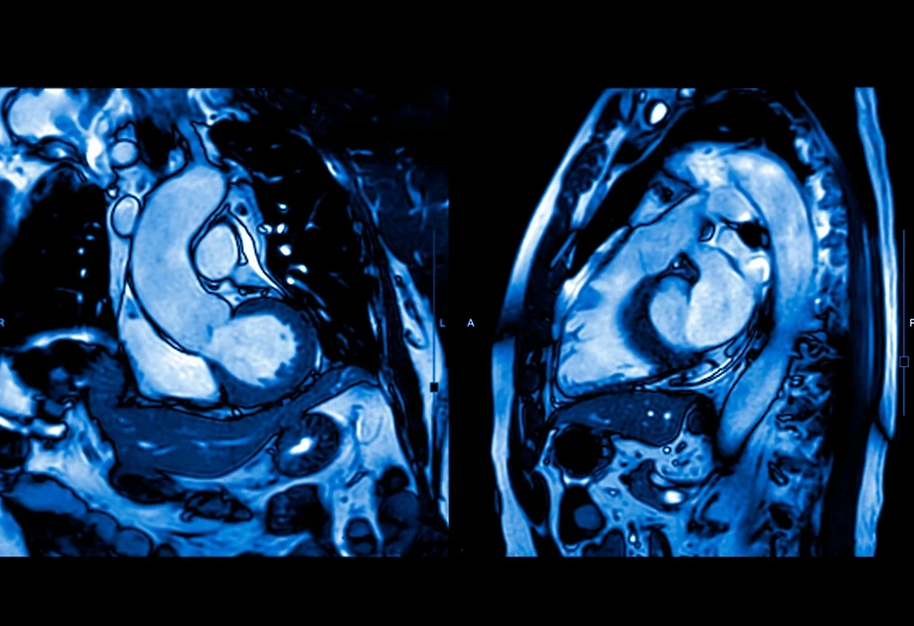
A cardiac MRI is a study that allows for the acquisition of high-resolution images of different areas of the heart to precisely evaluate its function.
What Is a Magnetic Resonance Imaging?
Magnetic resonance imaging works by using magnetic fields and the emission of radio waves, which allows for the acquisition of detailed images without being invasive to the patient or using radiation.
A cardiac MRI is commonly used to assess the size, shape, and function of the heart and to detect different conditions such as coronary artery abnormalities, congenital heart problems, or conditions located in a heart muscle.

A cardiac MRI is a non-invasive imaging study for which the patient lies on a stretcher and enters a closed tunnel of the equipment. Electrodes are placed on the chest to synchronize the images with the heart rhythm, which is known as electrocardiographic gating. In some cases, an intravenous contrast medium may be administered.
The procedure usually lasts between 30 and 60 minutes, depending on the type of study and the protocol used.
Cardiac MRI Protocol
The cardiac MRI protocol refers to the set of specific sequences and techniques used during the study to respond to a specific clinical suspicion.
Therefore, there is no single universal protocol, as it is adapted according to the condition to be evaluated, such as ischemic heart disease, cardiomyopathies, or congenital heart diseases, to name a few examples.
Almost all studies include functional images that allow for the observation of the heart’s movement in real-time and the evaluation of ventricular contractility.
In addition to this functional analysis, the protocol usually includes tissue characterization sequences, such as T1 and T2-weighted images, parametric maps, and late gadolinium enhancement. These techniques are fundamental for detecting inflammation, edema, fibrosis, or iron overload in the myocardium.
Each protocol is designed to obtain the maximum diagnostic information with the least possible discomfort for the patient.
Interpreting a Cardiac MRI
The interpretation of a cardiac MRI requires a detailed analysis by a medical specialist in cardiovascular imaging.
It begins with the evaluation of the moving images, where the systolic and diastolic function of the ventricles is analyzed, and volumes, ejection fraction, myocardial mass, and abnormalities in wall movement are calculated.
Subsequently, the tissue characterization sequences are examined to detect signs of edema, fibrosis, fatty infiltration, iron overload, or necrosis.
These findings are interpreted in the clinical context of each patient, as many imaging patterns are characteristic of specific conditions.
At the Cardiovascular Diagnostics area at ABC Medical Center, we can provide you with specialized care. Contact us!
Fuentes:
How can we help you?

Ricardo Ostos
Content CreatorRicardo can convey complex medical information in an accessible and friendly way so that all of our patients can understand and benefit from it. In addition, he has an empathetic approach, offering information and practical advice that really makes a difference in people's lives. #lifebringsustogether.
Learn more about Ricardo on LinkedIn
Pay in interest-free monthly installments in Specialty Centers, Check Ups, Diagnostic Tests, and Hospitalization
Get from 3 to 9 interest-free installments* with American Express or 6 installments* when paying with Banamex, BBVA, HSBC, Santander or 12 installments*
when paying with Banamex.
Privacy Overview
Error: Contact form not found.
We help you
Send us your request and we will forward it to our specialists. We will get in touch with you very soon.
If you have preferred times to receive our call, please indicate them in your message.
Thank you for contacting us!
Interest-free
months in:
Interest-free
months in:
Specialty Centers
Diagnostic Studies
Check-ups
Hospitalization1
Choose from3 to 9 months when paying with American Express cards 2. Or
6 months when paying with your credit card3 Banamex, BBVA Bancomer, HSBC, Santander.
Or 12 months exclusively when paying with Banamex3
Valid until December 31, 2025. Promotions not cumulative. Subject to restrictions 1. In hospitalization, medical fees are not included. 2. Minimum amount: $1,500 for 3 to 6 months and $3,000 for 7 to 9 months 3. Minimum amount $1,500. (Cards issued abroad are not eligible).
Comparison of COVID-19 vaccines
Pfizer-
BioNTech
Pfizer-BioNTech
What is its effectiveness and what does it refer to?
Vaccine type: mRNA
Effectiveness: 95% after the second dose in the prevention of symptomatic COVID-19.
No Does not contain egg, latex, or preservatives.
How many doses are needed?
Two doses are needed, at least 21 days apart (or up to six weeks apart, if necessary).
Who should or shouldn’t get the vaccine?
People who should receive the vaccine are those over 16 years old.
People who should not receive the vaccine are those who have a history of anaphylactic shock (severe allergy) or who are allergic to any component of this vaccine such as polyethylene glycol (PEG) or polysorbate.
What are the possible side effects of the vaccine?
Pain where the injection was given, fatigue, headache, muscle pain, chills, joint pain, fever, nausea, malaise, and swollen lymph nodes.
How long will it take for me to be protected and what does it protect me from?
After 14 days of having the complete scheme (after the administration of the 2nd dose), the protection period is still under study. It protects us from serious COVID-19 or requiring hospitalization.
Moderna
What is its effectiveness and what does it refer to?
Vaccine type: mRNA
Effectiveness: 94.5% after the second dose in the prevention of symptomatic COVID-19.
Does not contain egg, latex, or preservatives.
How many doses are needed?
Two doses are needed, at least 28 days apart (or up to six weeks apart, if necessary).
Who should or shouldn’t get the vaccine?
People who should receive the vaccine are those over 18 years old.
People who should not receive the vaccine are those who have a history of anaphylactic shock (severe allergy) or who are allergic to any component of this vaccine.
What are the possible side effects of the vaccine?
Pain where the injection was given, fatigue, headache, muscle pain, chills, joint pain, fever, nausea, and swollen lymph nodes in the arm in which you received the injection.
How long will it take for me to be protected and what does it protect me from?
After 14 days of having the complete scheme (after the administration of the 2nd dose), the protection period is still under study. It protects us from serious COVID-19 or requiring hospitalization.
Janssen/
Johnson
& Johnson
Janssen/ Johnson & Johnson
What is its effectiveness and what does it refer to?
Vector-based vaccine.
Effectiveness: 72.0% in the prevention of symptomatic COVID-19.
85% in the prevention of severe COVID-19.
Does not contain egg, latex, or preservatives./strong>
How many doses are needed?
Only one dose in needed.
Who should or shouldn’t get the vaccine?
People who should receive the vaccine are those over 18 years old.
People who should not receive the vaccine are those who have a history of anaphylactic shock (severe allergy) or who are allergic to any component of this vaccine.
What are the possible side effects of the vaccine?
Pain where the injection was given, headache, fatigue, muscle pain, chills, fever, and nausea.
How long will it take for me to be protected and what does it protect me from?
After 28 days of having the complete scheme (the last dose applied), the protection period is still under study. It protects us from 85% serious COVID-19 or requiring hospitalization.
AstraZeneca
and
Oxford
University
AstraZeneca and Oxford University
What is its effectiveness and what does it refer to?
Adenovirus vector-based vaccine.
Effectiveness: 82% after the second dose in the prevention of symptomatic COVID-19.
How many doses are needed?
Two doses are needed, at least 56 days apart (or up to 84 days apart, if necessary).
Who should or shouldn’t get the vaccine?
People who should receive the vaccine are those over 18 years old.
People who should not receive the vaccine are those who have a history of anaphylactic shock (severe allergy) or who are allergic to any component of this vaccine.
What are the possible side effects of the vaccine?
Pain where the injection was given, fatigue, headache, myalgia, arthralgia, and fever, which were mild to moderate in intensity and disappeared within 48 hours of vaccination.
How long will it take for me to be protected and what does it protect me from?
After 14 days of having the complete scheme (after the administration of the 2nd dose), the protection period is still under study. It protects us from serious COVID-19 or requiring hospitalization.
Sputnik V
What is its effectiveness and what does it refer to?
Adenovirus vector-based vaccine.
Effectiveness: 92% after the second dose in the prevention of symptomatic COVID-19.
How many doses are needed?
Two doses are needed, at least 21 days apart (or up to six weeks apart, if necessary).
Who should or shouldn’t get the vaccine?
People who should receive the vaccine are those over 18 years old.
People who should not receive the vaccine are those who have a history of anaphylactic shock (severe allergy) or who are allergic to any component of this vaccine.
What are the possible side effects of the vaccine?
Pain where the injection was given, fatigue, headache, myalgia, arthralgia, and fever, which were mild to moderate in intensity and disappeared within 48 hours of vaccination.
How long will it take for me to be protected and what does it protect me from?
After 14 days of having the complete scheme (after the administration of the 2nd dose), the protection period is still under study. It protects us from serious COVID-19 or requiring hospitalization.
Anti-Herpes Zoster
Herpes zoster is a painful, burning rash. It usually appears on one part of the body and can last for several weeks. It can cause long-lasting severe pain and scarring. Bacterial skin infections, weakness, muscle paralysis, hearing or vision loss may occur less frequently. Herpes zoster is caused by the same virus that causes chickenpox. After you have had chickenpox, the virus that caused it remains in the body of nerve cells. Sometimes after many years, the virus becomes active again and causes herpes zoster.
Vaccination is indicated in the following cases:
- Immunization of patients from 50 years of age for the prevention of herpes zoster and post-herpetic neuralgia (PHN), reduce pain associated with acute or chronic herpes zoster.
Scheme type:
- Single dose.
Rabies
Human rabies is a viral disease transmitted by the bite of an infected animal. It is characterized by acute encephalomyelitis (an aggressive response of the immune system that destroys the myelin layer of the nerves and alters its function at the level of the brain or spinal cord).
Vaccination is indicated in the following cases:
- Prevention of rabies in subjects exposed to risk of contamination. It is recommended for certain international travelers, based on the occurrence of animal rabies in the destination country.
Scheme type:
There are two types.
1. Pre-exposure scheme, consists of three doses of rabies vaccine:
- First dose, on the chosen date.
- Second dose 7 days after the first dose.
- Third dose 28 days after the first dose.
2. Post-exposure scheme, people not vaccinated against rabies, consists of five doses of rabies vaccine.
- First dose, on the date indicated.
- Second dose 3 days after the first dose.
- Third dose 7 days after the first dose.
- Fourth dose 14 days after the first dose.
- Fifth dose 28 days after the first dose.
* If the individual continues to be at risk of exposure to the disease, revaccination should be considered.
Pneumococcal vaccines
Pneumococcal disease can cause serious infections in the lungs (pneumonia), the bloodstream (bacteremia), and the lining of the brain and spinal cord (meningitis).
Two vaccines help prevent pneumococcal disease:
- Pneumoconjugate 13 (pneumococcal conjugate vaccine)
- Pneumococcal 23 (pneumococcal polysaccharide vaccine)
Vaccination is indicated in the following cases:
- Active immunization for the prevention of invasive disease caused by Streptococcus pneumoniae in adults 65 years of age and older.
Scheme type:
- *Two pneumococcal vaccines are recommended for all adults 65 years of age or older.
*One dose of Pneumococcal 13 vaccine should be given first, followed by one dose of Pneumococcal 23 vaccine, depending on your age and health.
