What Is a Uterine Fibroid: A Complete Guide to Uterine Fibromatosis
What is a uterine fibroid? (also known as a uterine leiomyoma, fibroma, or myoma) is a common question in gynecological care. It refers to a type of benign tumor that forms in the smooth muscle tissue, particularly in the uterus. This medical condition is one of the leading reasons for gynecological consultations today. Although the word "tumor" can cause immediate concern, it is essential to stay calm and understand its nature in order to address it with the clarity it requires.
This type of growth is non-cancerous and is primarily composed of muscle cells and fibrous tissue. Fibroids are very common in women of childbearing age; in fact, it is estimated that up to 70% of women will develop at least one fibroid at some point in their lives. Throughout this article, we will explore their causes, the range of symptoms they can cause, their impact on key life stages such as pregnancy, and the medical options available today for their proper management and treatment.
What Is a Uterine Fibroid and Why Does It Develop?
Understanding reproductive health involves learning about conditions that, due to their high prevalence, affect a large portion of the female population. The uterus is a muscular organ designed to expand and house life; however, due to various biological stimuli, its own cells can proliferate in isolation, giving rise to these growths. Below, we break down this condition conceptually and examine the scientific factors that explain how it develops in the body.
Definition of Uterine Fibromatosis
The term fibroid refers to benign tumors composed of smooth muscle tissue. While smooth muscle tumors can occur in other parts of the body (where they are broadly termed leiomyomas), fibroids most commonly affect the uterus in women. When a woman has multiple benign growths in her reproductive tract, the condition is medically referred to as uterine fibromatosis (or uterine fibroids).
These masses of smooth muscle tissue can vary drastically in size, ranging from tiny growths as small as an apple seed to bulky masses that alter the external anatomy of the uterus—sometimes visually mimicking an advanced pregnancy. Despite their growth, the cells that make up a fibroid do not invade distant tissues nor do they become cancerous in the future, meaning they are strictly classified as benign lesions.
Causes: Why Do Fibroids Develop in the Uterus?
The development of fibroids is influenced by a combination of factors, including environmental, hormonal, and genetic influences—though the exact mechanism triggering their onset is not fully understood. Hormonal factors are closely tied to estrogen and progesterone, which play a crucial role in their development. In women, these hormones regulate the menstrual cycle and stimulate the growth of uterine muscle cells, where fibroids originate.
During a woman’s reproductive years, levels of these hormones remain elevated, explaining why fibroids tend to grow steadily during this period. Conversely, they typically shrink significantly or naturally calcify once a woman reaches menopause, a stage marked by a substantial drop in estrogen production.
Primary Risk Factors
Genetic predisposition is another major factor; women with a family history of fibroids face a much higher risk of developing them. If a mother or sister has experienced uterine fibroids, the likelihood of developing the condition increases up to three times compared to the general population.
Lifestyle and environmental factors have also been shown to play a role. Some of these include:
- Overweight or obesity: Excess adipose (fat) tissue produces additional forms of estrogen, overstimulating uterine muscle tissue.
- Alcohol consumption: Particularly beer intake has been linked to higher risk.
- Vitamin D deficiency.
- Dietary choices: A diet rich in red meat and low in green vegetables is statistically associated with a higher incidence of these benign tumors.
Types of Fibroids and Common Symptoms
The clinical presentation of this condition varies from patient to patient. The exact location of the growth within the layers of the uterine wall dictates both the symptoms experienced and the medical approach specialists will choose for management or removal.
Intramural, Subserosal, and Submucosal Fibroids
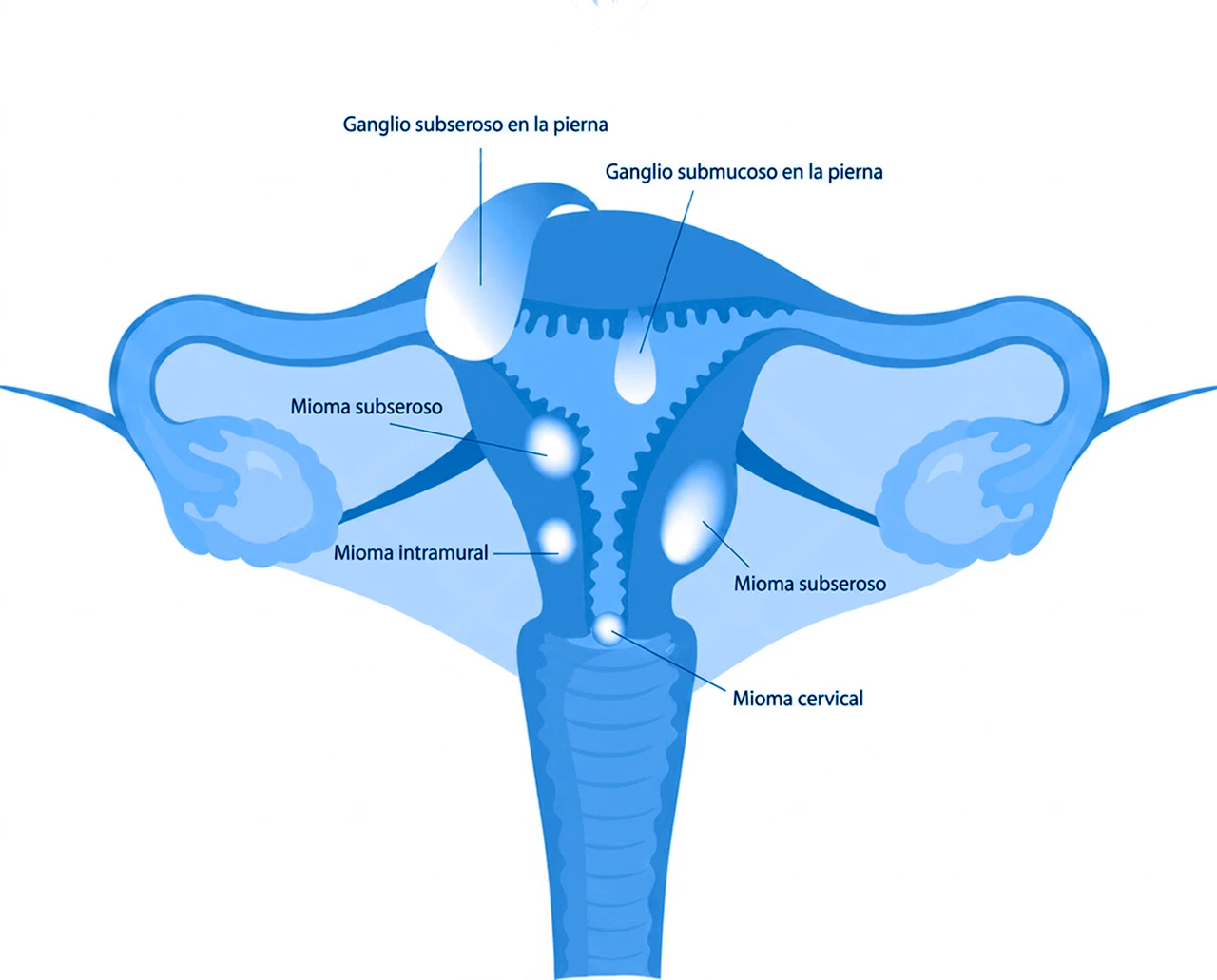
Fibroids can grow in different parts of the uterus, leading to a clear classification system for diagnosis:
- Intramural fibroids: Develop within the thick, muscular wall of the uterus. They are the most common type and, as they expand, cause the uterus to feel enlarged, often leading to dull aches and heavy periods.
- Subserosal fibroids: Grow on the outer layer of the uterus and expand into the pelvic cavity. Since they are outside the uterine cavity, they usually do not alter menstrual bleeding, but they can press against neighboring organs such as the bladder or bowel.
- Submucosal fibroids: Form just underneath the inner lining of the uterus (the endometrium). Although they account for the lowest percentage of cases, they cause the most severe issues with heavy bleeding and reproductive complications.
- Pedunculated fibroids: Attach to the uterus by a thin stem or stalk (pedicle), resembling a mushroom. They can originate as either subserosal or submucosal fibroids and cause acute pain if the stalk twists on its axis.
How Do I Know If I Have Fibroids? Warning Signs
Symptoms vary depending on the size, location, and number of fibroids. Uterine fibromatosis can also occur without presenting any noticeable symptoms. Among the most frequent signs are:
- Menorrhagia: Unusually heavy or prolonged menstrual bleeding.
- Intermenstrual bleeding: Spotting or bleeding between periods.
- Pelvic pain and pressure: Persistent fullness or discomfort in the lower abdomen.
- Urinary changes: Increased urinary frequency or difficulty emptying the bladder.
- Fertility issues: In certain cases, difficulty conceiving.
Prolonged, heavy bleeding should never be normalized, as it can lead over time to severe iron-deficiency anemia, characterized by chronic fatigue, pale skin, and shortness of breath during minimal physical effort. Pelvic pressure may also manifest as constant heaviness or persistent pain during sexual intercourse.
Fibroids During Pregnancy: Risks and Care
Pregnancy completely alters the hormonal environment of the female body, directly affecting any pre-existing uterine anomalies and requiring closer, more specialized obstetric monitoring.
Impact of Fibroids on Fertility
For many women, discovering a uterine mass raises immediate concerns about their ability to conceive. The reality is that most fibroids do not directly interfere with fertility. However, those classified as submucosal distort the inner shape of the endometrial cavity, which can prevent a fertilized egg from implanting properly or block the Fallopian tubes, hindering the sperm’s natural journey.
When severe fibromatosis distorts the overall anatomy of the organ, blood flow to the endometrium may be compromised. This can lower the success rates of assisted reproductive technology (ART) treatments unless corrective intervention is performed first to clear the uterine cavity.
Complications During Pregnancy and Childbirth
Developing or discovering fibroids during pregnancy is relatively common, often because a woman learns she has them during routine prenatal ultrasounds. In most cases, fibroids will not negatively impact the pregnancy or compromise the baby’s health.
However, in a small percentage of cases, complications can occur:
- Rapid fibroid growth: Driven by massive hormonal surges.
- Preterm labor: Caused by limited physical space inside the uterus.
- Fetal malposition: Such as breech positioning.
- Placental complications: Such as placental abruption (premature detachment).
- Increased likelihood of C-section: If the mass obstructs the birth canal.
Management during pregnancy is strictly limited to pain control, avoiding invasive surgeries that could put the gestation at risk.
Diagnosis and Treatment Options
Facing a fibroid diagnosis requires cutting-edge medical technology and a skilled team of gynecologists who can offer personalized treatment options, taking into account the patient’s age and future desire to preserve fertility.
Tests to Detect Uterine Fibroids
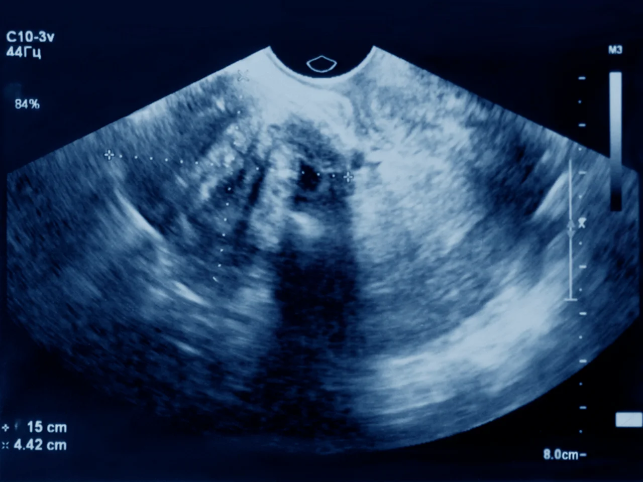
The diagnostic process typically begins during a routine gynecological exam through bimanual abdominal palpation, where the physician may detect an unusual enlargement of the uterus. To definitively confirm the diagnosis, a pelvic or transvaginal ultrasound is the imaging modality of choice, as it uses sound waves to quickly and painlessly map the exact size, number, and location of the fibroids.
In more complex scenarios or when delicate surgery is being planned, the specialist may request a magnetic resonance imaging (MRI) scan to obtain high-resolution 3D images, or a hysteroscopy, which involves inserting a tiny camera through the cervix to directly visualize the condition of the endometrial lining.
Medical Treatments and Symptom Management
Fibroids are a common condition—especially among women—and while they generally do not pose severe health risks, they can sometimes impact quality of life. For this reason, it is important to address them with healthcare professionals. The initial therapeutic approach for patients with mild symptoms is usually medical management, aimed at controlling pain and reducing heavy bleeding.
Common medical options include:
- Hormonal contraceptives: Oral pills, injections, or intrauterine devices (IUDs).
- Progesterone receptor modulators.
- Gonadotropin-releasing hormone (GnRH) agonists: These temporarily block estrogen production, inducing a temporary menopausal-like state that shrinks fibroids—often used as a preparatory step prior to surgery.
When Is Surgery Necessary?
Surgical intervention becomes the definitive option when symptoms significantly disrupt a patient’s daily life, if the fibroids grow rapidly, or if they impair the chances of a successful pregnancy. The classic surgical approach is a myomectomy, which involves removing only the fibroids while leaving the uterus intact—making it ideal for younger women who plan to have children in the future.
On the other hand, for women who have completed childbearing or who present with severe, uncontrollable uterine fibromatosis, a hysterectomy (total or partial removal of the uterus) provides a permanent resolution. Today, many of these procedures are performed using minimally invasive techniques such as laparoscopy or robotic surgery, ensuring rapid recovery times, reduced postoperative pain, and minimal cosmetic scarring.
Need Specialized Care?
The Women’s Health Center at ABC Medical Center is ready to provide you with specialized care. Contact us today!
Frequently Asked Questions
Fuentes: MedlinePlus, MSD Manual, Elsevier